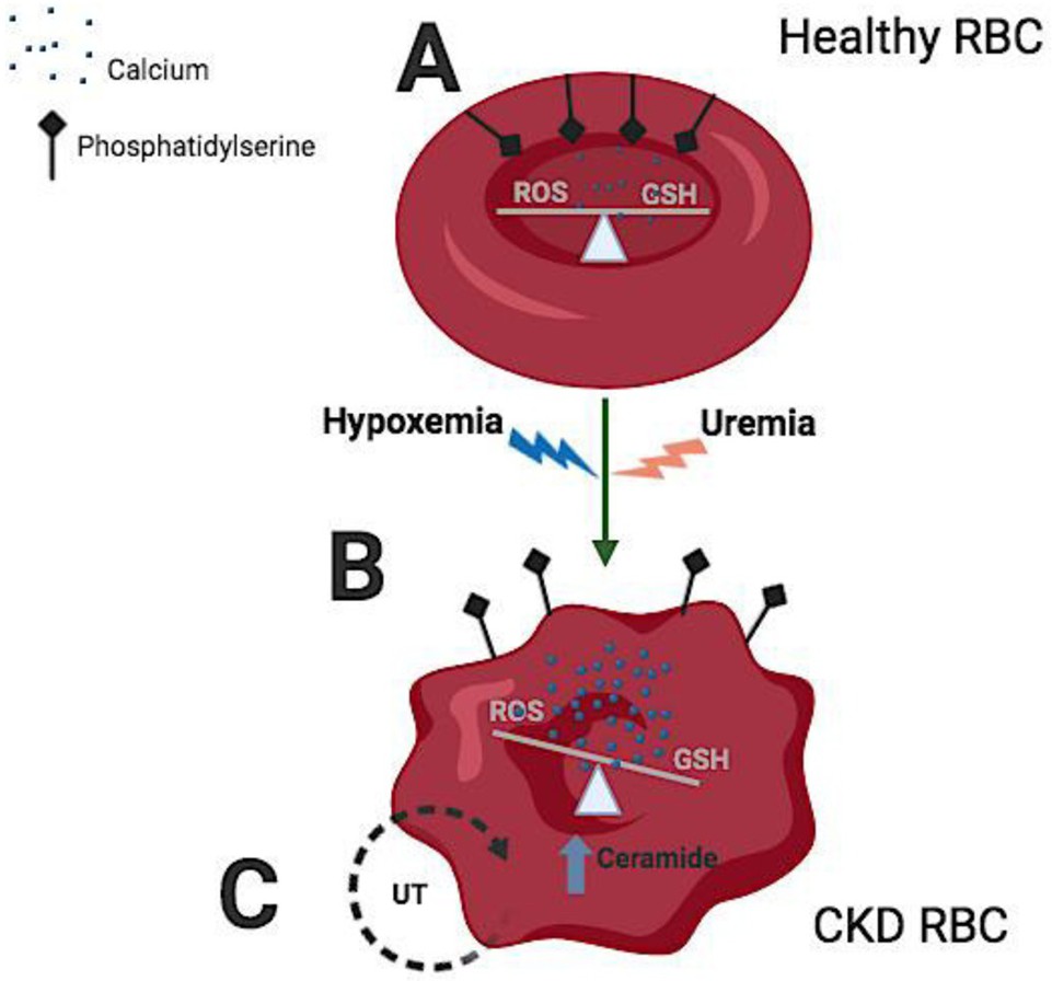
Fig. 7. Disturbance of red blood cell (RBC) homeostasis by uremia and hypoxemia. (A) Under normal conditions, healthy RBC maintain a normal calcium influx and a physiological balance between reduced glutathione (GSH) and reactive oxygen species (ROS). Under these circumstances, phosphatidylserine (PS) is located at the inner leaf of the RBC cell membrane and not exposed to the extracellular milieu. (B) In CKD, uremic toxins (UT), such as indoxyl sulfate (IS) accumulate; in addition, a low hemoglobin oxygen saturation (hypoxemia) is observed in a number of patients. These conditions in CKD RBC, favor an increased calcium influx and consequently a rise in intracellular calcium concentration. With the cellular redox state-maintained, the amount of ROS generation increases and GSH recycling is impaired. When the redox balance is disturbed, high ROS levels can disrupt the normal cellular machinery, eventually triggering the translocation of PS to the RBC surface, a signal that initiates RBC death (eryptosis). (C) Undertaking further study are necessary to understand 1. levels of ceramide formation under uremic and hypoxemic conditions, and 2. the impact of blocking the transport of the uremic toxins into RBC via organic anion transporter 2 (OAT2) on cell homeostasis. Fig. created using BioRender (https://app.biorender.com).